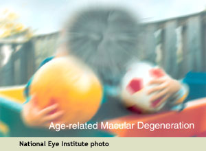
New research from University College London has challenged the traditional images that are used to show the visual symptoms associated with dry age-related macular degeneration (AMD), particularly the image at left from the United States National Eye Institute.
The researchers suggest that no one image can describe the condition and that “it may be more appropriate to develop a series of images or a dynamic representation, perhaps a series of movies or digital media, to more accurately depict vision in dry AMD. A more realistic description is important for raising awareness of the condition and educating patients.”
From the Journal Ophthalmic & Physiological Optics
This new research about macular degeneration imagery, titled Seeing it differently: self-reported description of vision loss in dry age-related macular degeneration, has been published online as an open-source article in the November 23, 2017 edition of Ophthalmic & Physiological Optics. The journal publishes original research on the development, use, and restoration of vision.
The authors are Deanna J. Taylor, Laura A. Edwards, Alison M. Binns, and David P. Crabb, from the Division of Optometry and Visual Science, School of Health Sciences, City, University of London, United Kingdom.
About the Macular Degeneration Imagery Research
Edited and excerpted from Images used to educate public about leading cause of blindness not realistic, says study, via Medical Xpress:
Images used to educate the public about vision loss due to the leading causes of blindness are not [realistic], according to a new study. The researchers found that the image commonly used to represent … age-related macular degeneration (AMD) … did not provide a realistic representation of people’s experiences.
When people with the condition were asked about how their vision looks, it was found that they instead provided a wide variety of descriptions – ranging from blurring, to distortion and missing parts of the image – which were far more complete and varied than those implied by these existing images, which just show a patch of distortion or blackness in central vision surrounded by a clear periphery.
As a result, the researchers call for the development of more realistic images of the visual symptoms of AMD for patient and public education which encompass the wide range of descriptions patients with the conditions use to describe the condition.
These descriptions could be used to educate people about the range of possible symptoms of dry AMD and would enable optometry professionals to better understand how the view of AMD through the patient’s eyes, leading to better recognition of symptoms for people with and without the condition.
The researchers found that people with dry AMD use a wide range of descriptors for their visual experience, with the most frequently reported descriptor “blur” (13 participants), followed by “missing” (10 participants) and “distortion” (seven participants). Furthermore, the visual symptoms of dry AMD as portrayed by commonly shown images were not the experience of most people in this study.
Said lead author Deanna Taylor, “We hope that through the development of new, more nuanced and realistic images, better education on AMD can be provided. This is especially important considering how the number of people with the condition is set to continue to rise in the UK and around the world over the coming decades.”
What Images Are Used to Show Vision Loss from Macular Degeneration?
This is the image developed by the National Eye Institute and used by many organizations and websites:
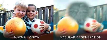
Here are some of the macular degeneration images we use on VisionAware:
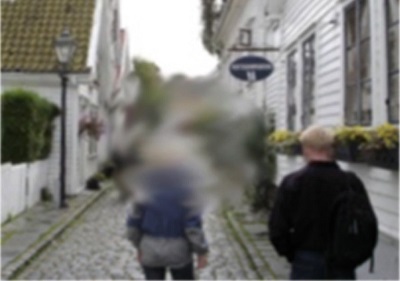
A simulation of central visual field loss from macular degeneration
Henry Ford Center for Vision Rehabilitation and Research
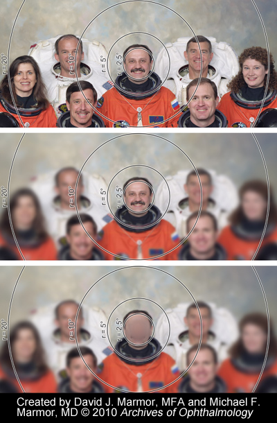
Top: How a camera would record a group of people
Middle: How a person with full vision would see it
Bottom: How a person with AMD would see it
What Is Age-Related Macular Degeneration?
Age-related macular degeneration (AMD) is a gradual, progressive, painless deterioration of the macula, the small sensitive area in the center of the retina that provides clear central vision. Damage to the macula impairs the central (or “detail”) vision that helps with essential everyday activities, such as reading, preparing meals, watching television, playing card and board games, and sewing.
AMD is the leading cause of vision loss for people aged 60 and older in the United States. According to the American Academy of Ophthalmology, 10-15 million people have AMD and about 90% of people who are affected have the “dry” type of AMD.
About Dry Macular Degeneration
The dry (also called atrophic) type of AMD affects approximately 80-90% of individuals with AMD. Its cause is unknown, it tends to progress slowly, and there is not – as of yet – an approved treatment or cure. “Atrophy” refers to the degeneration of cells in a portion of the body; in this case, the cell degeneration occurs in the retina.
In dry age-related macular degeneration, small white or yellowish deposits, called drusen, form on the retina, in the macula, causing it to deteriorate or degenerate over time.
A retina with drusen
Drusen are the hallmark of dry AMD. These small yellow deposits beneath the retina are a buildup of waste materials, composed of cholesterol, protein, and fats. Typically, when drusen first form, they do not cause vision loss. However, they are a risk factor for progressing to vision loss.
You can read more about macular degeneration at Age-Related Macular Degeneration at VisionAware.
More About the Study from Ophthalmic & Physiological Optics
Edited and excerpted from the study Abstract and Discussion:
Purpose: A realistic description of visual symptoms associated with dry age-related macular degeneration (AMD) is important for raising awareness of the condition and educating patients. This study aimed to develop a set of descriptors for dry AMD and examine the realism of images currently and frequently used to show visual symptoms of the condition.
Methods: Volunteers with dry AMD with a range of disease severity were given an eye examination and were asked to describe visual symptoms of their condition in a conversational interview. Participants were also asked to comment on a photograph typically used to portray the visual symptoms of AMD. Interviews were audio recorded, transcribed and subjected to content analysis.
Results: Twenty-nine participants were interviewed. Median age was 75 years. Median binocular visual acuity (VA) and contrast sensitivity (CS) was 0.2 and 1.65 respectively. Three, 17 and nine patients had early, intermediate and late AMD, respectively. The most frequently reported descriptor group was blur (13) followed by missing (10) and distortion (7). We chose the most popular image used to portray the visual symptoms of dry AMD based on an internet search and showed this to 21 participants. Sixteen participants, including three out of the seven people with [late AMD], rejected the realism of the image.
Conclusions: People with dry AMD use a wide range of descriptors for their visual experience. Visual symptoms of dry AMD as portrayed by commonly shown images were not the experience of most people in this study.
Discussion: Images showing “a patch of distortion” or “blackness” in central vision surrounded by a clear periphery are frequently used illustrators of vision with AMD. Our survey of a sample of images from an internet search supports this observation – three quarters of images showed virtually the same basic representation. However, only a small number of our sample of people with dry AMD reported this to be an accurate depiction of their visual experience and this was a key finding from our study.
Most people in our study did not think these images represented their visual symptoms. There was no strong evidence for this depiction representing visual symptoms for those with advanced dry AMD in the better-seeing eye either: only one person out of seven … stated that it was clearly representative of their visual symptoms.
Our main findings are important for several reasons:
First, the images we [examined] in this study are designed to educate the public about AMD and we have shown they are not fit for this purpose.
Second, the images could be misinterpreted to be a sign of early visual changes in AMD but this clearly does not fit with the experience of people with early or intermediate AMD in our sample.
Third, the visual symptoms experienced by most people with AMD are likely more subtle and less simplistic than those depicted in the images; this could have [consequences] for individuals about misunderstanding the severity of their own condition and may in turn affect adherence to management strategies such as self-monitoring of vision and lifestyle changes to minimize risk of disease progression.
Given the [wide variation] of the descriptors of visual symptoms reported in our study, it is perhaps unlikely that vision in dry AMD can be encompassed by a single image. It may be more appropriate to develop a series of images or a dynamic representation, perhaps a series of movies or digital media, to more accurately depict vision in dry AMD. Future studies might build on this idea.