A new study has revealed that African-Americans with diabetes have higher rates of vision loss from diabetic macular edema (explained below) compared with other ethnic and racial groups – and inconsistent access to eye care and eye examinations is a likely contributor to this disparity.
From JAMA Ophthalmology
The research, entitled Prevalence of and Risk Factors for Diabetic Macular Edema in the United States , was published in the November 2014 edition of JAMA Ophthalmology, an international peer-reviewed journal published monthly by the American Medical Association.
The authors are Rohit Varma, MD, MPH; Neil M. Bressler, MD; Quan V. Doan, PharmD; Michelle Gleeson, PhD; Mark Danese, PhD; Julie K. Bower, PhD; Elizabeth Selvin, PhD; Chantal Dolan, PhD; Jennifer Fine, PhD; Shoshana Colman, PhD; and Adam Turpcu, PhD, who represent the following institutions: Keck School of Medicine, University of Southern California, Los Angeles; Johns Hopkins University School of Medicine, Baltimore, Maryland; Outcomes Insights, Inc., Westlake Village, California; The Ohio State University College of Public Health, Columbus; and Genentech, Inc., South San Francisco, California.
About the Research
Excerpted from Vision loss more likely for African Americans with diabetes, via Futurity Research News:
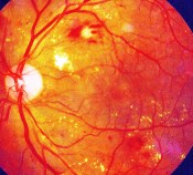
Diabetic macular edema (DME), one of the leading causes of blindness in diabetic patients in the United States, occurs when fluid and protein accumulates on the macula of the eye, which is part of the retina, causing it to thicken and swell. Central vision is affected and, if left untreated, can lead to slight blurring or even blindness.
“We were surprised that our research showed that African Americans have the highest rates of DME, when Hispanics tend to have the highest prevalence of diabetes,” said Rohit Varma, professor and chair of ophthalmology at University of Southern California (USC) and director of the USC Eye Institute.
“There is not enough vision screening for DME among diabetics, yet there are much better therapies available that are covered by insurance. We hope that our research will help those in the position to influence policy to get a better handle on costs and where the need for treatment is the greatest.”
For the study, researchers used the National Health and Nutrition Examination Survey (NHANES) database, a national data set measuring the health and nutritional status of American adults and children. As part of NHANES, subjects undergo a physical exam that includes photos of their retinas, which Varma’s team reviewed to determine the prevalence of DME.
Clinicians should assess diabetes patients, especially those who are African American or Hispanic, more closely for vision loss, Varma says. Also, patients should do everything they can to control their glucose and monitor their own vision. Varma says he will next examine barriers African Americans face concerning access to eye care.
About Diabetic Macular Edema
Diabetic macular edema [edema = a swelling or accumulation of fluid] (DME) can occur in people with diabetes when retinal blood vessels begin to leak into the macula, the part of the eye responsible for detailed central vision. These leakages cause the macula to thicken and swell, which, in turn, creates a progressive distortion of central vision.
Although this swelling does not always lead to severe vision loss or blindness, it can cause a significant loss of central, or detail, vision, and is the primary cause of vision loss in people with diabetic retinopathy.
Treatments for Diabetic Eye Disease
The first step in any treatment for diabetic eye disease is to maintain blood glucose, blood pressure, and blood cholesterol levels as close to normal as possible.
Treatment of diabetic macular edema has evolved a great deal in the last five to ten years, and is based on the severity of the edema. At present, there are three options:
- laser treatment
- Avastin, Lucentis, or Eylea injection
- intravitreal steroids
Laser Treatment
This technique is used by retinal surgeons to treat a number of eye conditions, one of which is diabetic eye disease. A beam of high-intensity light is directed into the eye to seal off leaking blood vessels and prevent additional blood and fluid from leaking into the vitreous. The doctor administers eye drops to dilate the pupil and numb the eye before treatment begins.
Because lasers cannot restore lost vision, it is critical to maintain regular eye examinations so that treatment can be initiated as soon as diabetic eye changes are detected. There are two types of laser treatments for diabetic eye disease:
- Focal laser treatment, also called photocoagulation: The retina is treated to stop or slow the leakage of blood and fluid from abnormal blood vessels within the eye. Focal laser, however, can also destroy surrounding healthy retinal tissue as it seals the leakage from abnormal blood vessel growth; therefore, it is not used on blood vessels directly under the macula, the center of the retina.
- Scatter laser treatment, also called panretinal [i.e., encompassing the entire retina] photocoagulation: The areas of the retina away from the macula are treated to shrink abnormal blood vessels.
Avastin, Lucentis, or Eylea Injections
In diabetic eye disease, abnormal blood vessels develop that can break, bleed, and leak fluid. If left untreated, these damaged blood vessels can result in a rapid and severe loss of vision. The most effective treatments to date for this blood vessel damage are the anti-angiogenic drugs Avastin, Lucentis, and Eylea.
Angiogenesis is a term used to describe the growth of new blood vessels and plays a crucial role in the normal development of body organs and tissue. Sometimes, however, excessive and abnormal blood vessel development can occur in diseases such as cancer (tumor growth) and diabetic eye disease (retinal and macular bleeding).
Substances that stop the growth of these excessive blood vessels are called anti-angiogenic (anti = against; angio = vessel; genic = development), and anti-neovascular (anti = against; neo = new; vascular = blood vessels).
The focus of current anti-angiogenic drug treatments for diabetic eye disease is to reduce the level of a particular protein, called vascular endothelial growth factor or VEGF, that stimulates abnormal blood vessel growth in the retina; thus, these drugs are classified as anti-VEGF treatments. At present, Avastin, Lucentis, and Eylea are administered by injection directly into the eye after the surface has been numbed.
These drugs are powerful. The abnormal vessels will disappear within 24 to 48 hours; however, the vessels are not gone forever. They will come back, since the effect of the drug will wear off. The half-life of the drugs in the eye is about four to six weeks. Treating edema with these drugs requires frequent treatment.
Intravitreal Steroids
Kenalog or triamcinolone provide good control of the swelling caused by diabetic macular edema. Like Avastin, Lucentis, and Eylea, the steroid is injected into the eye; the swelling will disappear within 24 hours.
Steroids do, however, have some side effects. One in ten patients develop glaucoma from the steroid shot. Steroids can also cause cataracts. Nevertheless, steroid treatment can be a useful tool and can be combined with laser. This combination can eliminate the swelling and “dry up” areas of leaking blood vessels.
You can read more about treatments at What Treatments Are Available for Diabetic Eye Disease? by Lori Coors, M.D. at the VisionAware website.
More about the Study from JAMA Ophthalmology
From the article abstract:
Importance: Diabetic macular edema (DME) is a leading cause of vision loss in persons with diabetes mellitus. Although there are national estimates for the prevalence of diabetic retinopathy and its risk factors among persons with diabetes, to our knowledge, no comparable estimates are available for DME specifically.
Objective: To estimate the prevalence of DME in the US population and to identify associated risk factors.
Design, Setting, and Participants: A cross-sectional analysis of 1,038 participants aged 40 years or older with diabetes and valid fundus [i.e., retinal] photographs in the 2005 to 2008 National Health and Nutrition Examination Survey.
[Editor’s note: A cross-sectional study analyzes and studies a population of subjects at one specific point in time, rather than over a longer, or more extended, period of time.]
Main Outcomes and Measures: The overall prevalence of DME and its prevalence according to age, race/ethnicity, and sex.
Results: Of the 1,038 persons with diabetes analyzed for this study, 55 had DME, for an overall weighted prevalence of 3.8% or approximately 746,000 persons in the US 2010 population aged 40 years or older. We identified no differences in the prevalence of DME by age or sex.
Multivariable logistic regression analysis showed that the odds of having DME were higher for non-Hispanic blacks than for non-Hispanic whites. Elevated levels of glycosylated hemoglobin A1c for each 1% and longer duration of diabetes (greater than or equal to 10 years vs. less than 10 years) were also associated with DME prevalence.
[Editor’s note: The glycated hemoglobin (A1c) test, which can be done without fasting, measures the person’s average blood glucose control for the past 2 to 3 months. A level between 5.7 and 6.4 suggests an increased risk of developing diabetes; a level greater than or equal to 6.5 indicates diabetes.]
Conclusions and Relevance: These results suggest a greater burden of DME among non-Hispanic blacks, individuals with high levels of hemoglobin A1c, and those with longer duration of diabetes. Given recent treatment advances in reducing vision loss and preserving vision in persons with DME, it is imperative that all persons with diabetes receive early screening; this recommendation is even more important for those at higher risk for DME.